

Translate this page into:
A rare incidental case of an accessory fallopian tube
-
Received: ,
Accepted: ,
This is an open access journal, and articles are distributed under the terms of the Creative Commons Attribution-NonCommercial-ShareAlike 4.0 License, which allows others to remix, tweak, and build upon the work non-commercially, as long as appropriate credit is given and the new creations are licensed under the identical terms.
This article was originally published by Wolters Kluwer - Medknow and was migrated to Scientific Scholar after the change of Publisher.
Abstract
Developmental anomalies of the müllerian duct system represent one of the most fascinating disorders that obstetricians and gynecologists encounter as the müllerian ducts are the primordial anlage of the female reproductive tract. Accessory fallopian tube is one such rare anatomical müllerian duct error that has been occasionally reported in the literature. Due to the limited data available and lack of awareness about this entity, it is often overlooked. This report describes one such rare incidental case of an accessory fallopian tube in a 35-year-old female who was diagnosed with right ruptured tubal pregnancy. The patient was about six weeks pregnant and presented with a severe lower abdominal pain. Transvaginal sonography showed that the right fallopian tube contained a gestational sac with a yolk sac and her urine pregnancy test was positive, so a diagnosis of a right ruptured tubal pregnancy was made. Laparotomy and right salpingectomy were performed. Histopathological examination revealed right ruptured tubal pregnancy with a coexisting accessory fallopian tube. In conclusion, an early identification and prompt intervention are paramount for treating such an anomaly as it can have many gynecological detrimental implications.
Keywords
Accessory fallopian tube
ectopic pregnancy
infertility
mullerian duct anomalies

INTRODUCTION
The female reproductive tract develops from a pair of müllerian ducts that undergo organogenesis, fusion and resorption in utero to give rise to the uterus, fallopian tubes, cervix, and upper two-thirds of the vagina. Therefore, any interruption in the müllerian ducts development during embryogenesis can result in formation of müllerian duct anomalies (MDAs), which are a wide and a complex spectrum of congenital abnormalities that are often associated with renal as well as axial skeletal anomalies or can be a part of multiple malformation syndrome and may cause numerous other gynecological complications.[1]
The reported incidence of MDAs in routine clinical practice has been documented as 0.1–0.5% in general and up to 6% in patients with infertility.[2] Most of them are encountered in uterus followed by vagina and cervix. Isolated congenital anomalies of the fallopian tubes are uncommon and are often overlooked owing to the low index of suspicion. Various anatomical variants such as phimoses, accessory tubes and tubal ostia, sacculations, complete absence/partial atresia or segmental deletion of different regions of the tube and fimbrial agglutinations have been seldom documented in the literature.[3] These variations are harbinger of infertility, ectopic pregnancy, hydrosalpinx, pyosalpinx, cystic swelling, pelvic inflammatory diseases, endometriosis and torsion. Therefore, early identification and correction of these tubal subtle lesions may be beneficial in preventing infertility and improving the pregnancy rates. Herein is described an extremely rare case of clinically unsuspected accessory fallopian tube which was incidentally diagnosed during routine histopathological examination of a 35-year-old female who had undergone right salpingectomy for right ruptured tubal pregnancy.
CASE REPORT
A 35-year-old female, G2P1, presented to the gynecological outpatient department with a sudden onset of severe lower abdominal pain of 24-hours duration. She was about six weeks pregnant and a urine pregnancy test was positive four days prior to her admission. There was no complaint of vaginal bleeding. She had one previous normal pregnancy and a vaginal delivery of a healthy boy, two years back. Her last menstrual period was on 2 March 2019. Her menstrual cycle was regular, lasting five days, every 28 days with an average flow. Her past medical history for any major disease or prior surgeries as well as family history was non-contributory. On general physical examination, she was thin built and anemic. Her pulse rate was 116 per minute and regular. Her blood pressure was 100/80 mm Hg. Her temperature was 36.8°C. Per abdomen examination revealed slight distention and mild tenderness in the lower abdomen with no evidence of ascites or any organomegaly. Per speculum examination showed a healthy vulva, vagina and urethra. Bimanual pelvic examination revealed cervical excitation as well as an anteverted, soft, normal-sized uterus with free bilateral fornices and without palpable adnexal masses. All other systemic examinations were within the normal limits. Her routine hematological investigations revealed microcytic hypochromic blood picture. Urine and blood cultures were negative. Kidney and liver function tests were normal. Serum antibodies to human immunodeficiency virus, hepatitis B surface antigen, syphillis were negative. X-ray chest was normal. Transvaginal sonography (TVS) showed an empty uterine cavity, normal ovaries and left fallopian tube with a large volume of complex free fluid with internal echogenicity in the Pouch of Douglas. However, the right fallopian tube contained a gestational sac with a yolk sac. A repeat urine pregnancy test was positive. On the basis of the history, clinical, laboratory and TVS findings, a diagnosis of a right ruptured tubal pregnancy was made. The patient was immediately taken up for laparotomy which was followed by right salpingectomy and the specimen was sent for histopathological examination. Grossly, a distended right fallopian tube measuring 3.5 × 2.5 × 1.5 cms in size was received. Its external surface showed congestion and a site of rupture. On further, careful inspection of the right fallopian tube, another hypoplastic fallopian tube with its own fimbria measuring 1 × 0.3 × 0.3 cms was seen attached to its ampullary segment [Figure 1]. On cut section, of the right main fallopian tube, blood clots along with gestational sac were seen. A probe was passed from the main tubal ostia into the lumen of the accessory tube; however it did not appear to be patent with the main tube. The cut section of the accessory tube was unremarkable. Microscopic sections of the right main fallopian tube exhibited a site of rupture along with a lumen lined by unremarkable tubal lining and comprising of extensive hemorrhage, numerous chorionic villi and trophoblastic cells. On the other hand, the sections from the accessory fallopian tube showed a fimbrial end and a stalk with tortuous vessels, fibrous tissue, and smooth muscle. The fimbrial end was open, and the lumen that penetrated down the stalk had typical tubal epithelial folds [Figure 2]. Based on the histopathological findings, a final diagnosis of right ruptured tubal pregnancy with a coexisting accessory fallopian tube was rendered. The patient underwent further vigilant inspection and ultrasonography of the abdomen as well as pelvis which however revealed no other genitourinary or pelvic anomaly. The postoperative recovery of the patient was uneventful. She was discharged home on the tenth postoperative day and is still under weekly follow-up.

- An accessory tube attached to the ampullary segment of the oviduct.
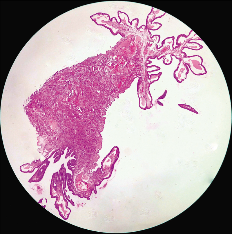
- Histopathological section exhibiting an open fimbrial end and a stalk with typical tubal epithelial folds in the lumen (H and E, x100).
DISCUSSION
The fallopian tube diseases account for about 25%–35% of all the female infertility cases, however, they are often under-estimated and misdiagnosed, thus leading to unwarranted diagnostic and therapeutic implications. In literature, a variety of non-neoplastic and neoplastic pathologies of the fallopian tubes exists, nevertheless amongst all it is the anatomical abnormalities of the fallopian tubes which are of great clinical significance as although they occur rarely but can lead to life-threatening consequences, if not timely intervened. One such extremely unusual developmental malformation is accessory fallopian tubes, which results from bifurcation of the cranial ends of the müllerian ducts, which normally develops into fallopian tubes. Their exact incidence is unknown, because most of the times the surgeons fail to notice this anomaly, as encountered in the present case too.
The review of the literature shows that, it was in the year 1894, that Kossman et al., were the first to describe the details of accessory tubes as well as of accessory ostia and mentioned their incidence to be in 4% to 10% of all women.[4] Later, in 1904, Macnaughton-Jones H. studied the relation between the accessory fallopian tubes, broad ligament cysts and hydrosalpinx.[5] In 1948, Gardner et al., reviewed this subject partially in their study.[6] This was followed by Zolcinski et al., in 1964, who found accessory tubes in nearly 5% of their operations and cesarean sections.[7] In the year 1982, Beyth et al., studied 200 abdominal operations and found the incidence of accessory tubes in their experience to be approximately 6%.[8] Similar incidence of accessory fallopian tubes as high as 6% was also mentioned by Coddington et al., in the year 1990.[9] Since then, according to the pertinent world literature, there are only a handful of accessory fallopian tubes cases which have been reported so far.[10,11,12,13,14,15]
Most of the patients harboring this rare anomaly are usually asymptomatic and are diagnosed incidentally on laparoscopy for some unrelated purpose. The present case was also discovered by chance on histopathological examination of the right salpingectomy specimen which was obtained after a ruptured right tubal pregnancy was detected clinically. Due the sporadicity of such cases and limited data available about this entity, the possibility of accessory fallopian tubes is often not kept which in turn causes various complications and can have a fatal outcome.
These accessory fallopian tubes are a major contributing factor of infertility and ectopic pregnancy as fimbria are capable of picking up ova even if they are not in their normal anatomical location, that is, close to the ovary. Also, the occlusion of a segment of the fallopian tube does not interfere with the ovum pickup mechanism by the fimbria. Therefore, in the presence of accessory tubes with fimbriated ends, ova may eventually be captured by the fimbria, instead of the fimbria of the main normal fallopian tube. Any ovum so captured will decrease the chances of an intrauterine pregnancy. Another concept is that of transperitoneal migration of sperm where the sperm that enter the peritoneal cavity through the main tubal ostium may migrate to the fimbria of the accessory tube and fertilize a captured ovum there, resulting in an ectopic pregnancy within the accessory tube, which is further more prone to excessive bleeding as well as requires an emergency surgical intercession. An additional important aspect of it is that in this era of medico-legal issues, it can be a potential cause for failure of postpartum sterilization procedure, which is actually worrisome to both the patient and the clinician dealing with such cases.[10] This anomaly can also be associated with other MDAs as well as renal malformations including agenesis, ectopia, hypoplasia, fusion, malrotation, and duplication.[1] Therefore, patients with such an anomaly should also be evaluated for any concomitant aberrations. The case presented was also screened for such abnormalities however, no such variations were found. Nevertheless, similar to this case, authors have documented and well-supported the fact that when congenital ampullary atresia occurs without synchronous MDAs, then the increased chances of having associated renal abnormalities are unlikely.[16] Other repercussions of the accessory fallopian tubes can be torsion, hydrosalpinx, pyosalpinx, cystic swelling and pelvic inflammatory diseases. Therefore, its early identification via pelvic ultrasonography, hysterosalpingography, or laparoscopy and preventive removal of such an anomaly microsurgically followed by histopathological confirmation should be considered so as to avoid any dreadful consequences.[10,13]
CONCLUSION
Accessory fallopian tubes are one of the rare MDAs which are under-reported due to the medical unfamiliarity and diagnostic subtleties associated with the condition. Nevertheless, this entity should always be kept in mind especially while dealing with the infertility cases. A high index of suspicion and meticulous systematic examination of the fallopian tubes during abdominal surgeries is important for its early recognition and to prevent many gynecological complications.
Financial support and sponsorship
Nil.
Conflicts of interest
There are no conflicts of interest.
REFERENCES
- Congenital absence of a part of the fallopian tube: a case report. Int J Reprod Contracept Obstet Gynecol. 2017;6:320-2.
- [Google Scholar]
- Clinical implications of accessory fallopian tube ostium in endometriosis and primary infertility. Womens Health (Lond). 2016;12:404-6.
- [Google Scholar]
- Accessory fallopian tubes and their relation to broad ligament cysts and hydrosalpinx. BJOG. 1904;6:212-5.
- [Google Scholar]
- Normal and cystic structures of the broad ligament. Am J Obstet Gynecol. 1948;55:917-39.
- [Google Scholar]
- Accessory tubes: A possible contributing factor in infertility. Fertil Steril. 1982;38:382-3.
- [Google Scholar]
- Accessory fallopian tube in an adolescent female: a case report. J Pediatr Adolesc Gynecol. 2009;22:e27-8.
- [Google Scholar]
- Isolated torsion of accessory fallopian tube in a young adolescent. J Pediatr Adolesc Gynecol. 2016;29:e57-8.
- [Google Scholar]
- Patient with three fallopian tubes at right adnexa. J Clin Diagn Res. 2017;11:QJ03-4.
- [Google Scholar]
- Fallopian tube duplication: a rare anomalistic case report with review of literature. AWCH. 2017;3:C11-2.
- [Google Scholar]
- Congenital interruption of the ampullary portion of the fallopian tube. Fertil Steril. 2006;85:1820-1.
- [Google Scholar]


