Translate this page into:
Maternal and neonatal outcome of surviving twin after single fetal demise at 25 weeks: A rare case report
Address for correspondence: Dr. Manisha Choudhary, 20/133, Kaveri Path, Mansarovar, Jaipur 302020, Rajasthan, India. E-mail: drmanishachowdhary@gmail.com
This is an open access journal, and articles are distributed under the terms of the Creative Commons Attribution-NonCommercial-ShareAlike 4.0 License, which allows others to remix, tweak, and build upon the work non-commercially, as long as appropriate credit is given and the new creations are licensed under the identical terms.
This article was originally published by Wolters Kluwer - Medknow and was migrated to Scientific Scholar after the change of Publisher.
Abstract
Single fetal demise in twin pregnancy in late second or third trimester is a complex clinical situation and the management may face a dilemma. This is a rare case report of continuing pregnancy for 9 weeks in an intrauterine insemination (IUI) conceived dichorionic diamniotic twin pregnancy with intrauterine fetal demise of one twin at 25 weeks of gestation. A gravida 2 abortion 1 presented to Jaipur fertility centre (Department of Reproductive Medicine & Medical Genetics, MGUMST) with unexplained infertility of 3 years. She conceived with IUI at first attempt. The routine antenatal care scan at 25 weeks revealed 25 weeks 3 days single live fetus and second fetus 21 weeks with absent fetal heart pulsation and features of hydrops. The patient was hospitalized for conservative management. Regular follow-up was performed with daily fetal movement count, weekly coagulation profile, and ultrasound for fetal well-being. Inj. betamethasone was given for lung maturation at 32 weeks of gestation. She underwent caesarean section at 34 completed weeks for the preterm premature rupture of the membrane, the outcome being first fetus 2.2 kg female with Apgar score 7/10 and second macerated female fetus of 840 g. Postoperative period was uneventful for both mother and newborn. The baby was on regular check-up under a neonatologist. Her growth and neurological development was optimum according to her age group as seen on the long-term follow-up for 1 year.
Keywords
Caesarean section
diamniotic dichorionic twin pregnancy
DIC
NICU
single fetal demise
INTRODUCTION
Multifetal pregnancies are associated with a higher risk of perinatal morbidity and mortality when compared with singleton pregnancies. The incidence of single fetal death in twin pregnancies is 2.5–5.0%[1] as compared to 0.3–0.6% in singletons.[2] The causes of single fetal demise in a twin pregnancy include twin–twin transfusion, placental insufficiency, intra uterine growth restriction related to pre-eclampsia, velamentous insertion of cord, cord stricture or true knot, cord around the neck, and congenital abnormalities.[3] Single fetal demise in a twin pregnancy before 17 weeks is usually uneventful. The death of a twin in late second or third trimester is a rare obstetric complication and a cause of great concern and psychological stress to both parents and obstetrician. This may increase the risk of preterm labor, pre-eclampsia, maternal disseminated intravascular coagulation (DIC), intra uterine growth restriction, neurological complication, or even death of surviving twin.
Chorionicity rather than zygosity determines the risk of complications in surviving twin. The prevalence of monochorionicity is up to 50–70%[4,5] in single fetal demise in twins. Assessment of chorionicity in early fetal scans in a twin pregnancy is mandatory. In monochorionic pregnancies, the risk of morbidity and mortality in surviving twin is 3- to 4-fold greater than in dichorionic pregnancies. In a study by Livnat et al.,[6] they described a 12% risk of co-twin death in monochorionic twins as compared to a 4% risk in dichorionic pregnancies.
The precise cause of poor perinatal outcome of the surviving co-twin in monochorionic pregnancies is unknown. The reported frequency of vascular connections in monochorionic placentas range from 85 to 98%.[7,8] Because of the presence of placental anastomoses, the death of one twin can cause an acute hypotension, anemia, and multiorgan ischemia in the co-twin thought to be a result of the acute shunting of blood to the demised fetus. The other mechanisms could be transchorionic embolization and coagulopathy. In a dichorionic pregnancy, these vascular anastomoses are not present, but the intrauterine environment that may have caused the initial fetal demise such as infection or maternal medical diseases may place the surviving twin at risk as well. Major morbidities are unlikely to occur in the surviving twin.
Maternal coagulopathy (DIC) is the most feared complication following single fetal demise which is reported to occur 3–5 weeks following demise. The release of fibrin and thromboplastin from dead tissues into maternal circulation is the probable mechanism of maternal DIC and can be fatal for both the mother and the fetus. So the continuation of pregnancy beyond 5 weeks is more risky and less reported.
Recent changes in technology and repeated prenatal ultrasound scan make it possible not only to diagnose this condition timely but also helps in close fetal surveillance during conservative management. The individual studies including data from case reports, follow-up of cohorts, and twin registries tend to have imprecise results as the event is uncommon. The absence of large-scale studies makes it difficult to advise the parents on the prognosis and optimal management but conservative management is preferred by most obstetricians.
CASE REPORT
A 28-year-old G2A1 presented to Jaipur fertility centre with secondary infertility of 3 years. On complete workup, the cause was attributed to unexplained infertility.
She conceived with intrauterine insemination (IUI) at first attempt. The protocol used was inj. Fostine 75 IU for 7 days followed by inj. Menogon 150 IU for 2 days. Following successful IUI, she had a twin pregnancy with last menstrual period − 13/12/15 and expected date of delivery − 20/09/16. Confirmation scan at 6 weeks of gestation showed dichorionic diamniotic twins. Her nuchal translucency (NT) scan at 11 weeks 5 days revealed NT − 1.3 and 1.4 mm, respectively with low lying placenta. Other routine investigations such as complete blood count (CBC), urine complete and microscopic tests, thyroid stimulating hormone, and glucose challenge test were within normal limit. Level 2 scan at 19 weeks 3 days was normal for both fetuses.
The routine antenatal care scan at 25 weeks revealed a 25 weeks 3 days single live fetus and second fetus 21 weeks with absent fetal heart pulsation and features of hydrops. The patient was hospitalized for observation. On general examination, she was hemodynamically stable, afebrile, and was normotensive. Other systemic examination revealed no abnormality. On per abdominal examination, fundal height was about 30 weeks pregnancy size with multiple fetal parts felt. The uterus was not tense and nontender. On auscultation, single fetal heart sound was 144 beats/min. There was no per vaginal bleeding or abnormal discharge.
Her investigations showed hemoglobin − 11.1 g/dl, total white blood cell count − 11,900/cumm, bleeding and clotting time being normal, prothrombin time (PT) − 14.0, inrnational normalized ratio (INR) − 1.27 s and activated partial thrombplastin time (APTT) − 41.4 s, serum fibrinogen − 457 mg/dl, and total platelet count − 2.9 lacs. Blood group was AB+ve. HBsAg, human immunodeficiency virus and venereal disease research laboratories were nonreactive. Her urine routine and c/s with high vaginal swab c/s were all within normal range. After admission, prophylactic broad-spectrum oral antibiotic was given for 1 week. Tab. Ecosprin 75 mg OD and inj. Proluton depot 250 mg i/m weekly continued till 32 weeks of gestation. 12 mg betamethasone two doses 24 h apart were given for lung maturation at 32 weeks of gestation.
Maternal monitoring included weekly CBC, DIC profile (PT, INR, APTT, and serum fibrinogen), fortnightly high vaginal swab and urine culture, and sensitivity. All were within normal limit till delivery. Fetal monitoring included weekly ultrasound (USG) for fetal well-being and nonstress test (NST) started at 32 weeks biweekly, which was all reactive.
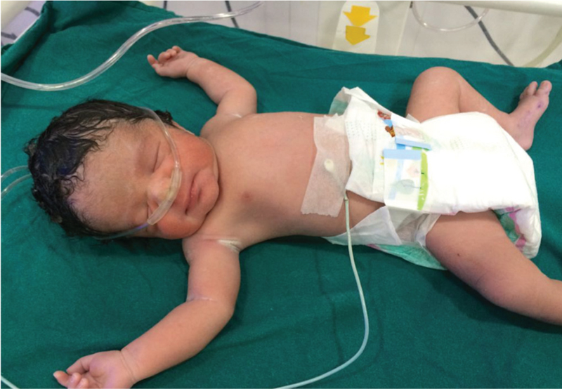
She underwent caesarean section at 34 completed weeks, after informed and neonatal intensive care unit (NICU) consent, the indication being preterm premature rupture of membrane. On the day of caesarean section, the patient complained of leaking p/v. On per speculum examination, frank leak was present and on per vaginal examination, os was closed and uneffaced. Investigations on the day of caesarean section were hemoglobin − 11.9 g/dl, total white blood cell count − 10,340/cumm of blood, bleeding and clotting time − normal, PT − 12.7, INR − 1.12 s and APTT − 29.8 s, serum fibrinogen − 635 mg/dl, platelet count − 2.7 lacs. The outcome was first fetus − 2.2 kg female [Figure 1] with Apgar score − 7/10. The baby was referred to NICU due to low birth weight and prematurity [Figure 2]. The second fetus was 840 g female, which was macerated. Intraoperative findings revealed dichorionic diamniotic sac with no sign of infection, growth, or other abnormalities in the placenta and cord of surviving twin. The dead fetus was in separate intact sac, and the cord was normal in length. There were no apparent gross abnormalities in the placenta and the cord with no retroplacental clots.

- Female 2.2 kg – the surviving co-twin

- Surviving twin in NICU (low birth weight and prematurity)
The placenta of the dead fetus was sent for histopathological examination [Figure 3] which revealed only necrotic tissue. The dead fetus was sent for autopsy, which showed no detectable cause for fetal demise. During postoperative period, injectable broad-spectrum antibiotics and analgesics were given to the mother. Both the mother and the baby were healthy and discharged on 7th postoperative day. The baby was on regular checkup under a neonatologist. Her growth and neurological development was optimum according to her age group as seen on long-term follow-up for 1 year.

- Healthy placenta of surviving co-twin. Macerated co-twin with its placenta
DISCUSSION
We are presenting this case report because it is rare of a kind and challenging to continue pregnancy for 9 weeks in a case of twin pregnancy with intrauterine fetal demise of one twin at 25 weeks period of gestation. It included vigilant maternal and fetal monitoring resulting in healthy fetus at 34 completed weeks of gestation. The incidence of twin pregnancy with a single fetal demise has been reported as 0.5–6.8% by Enbom,[9] 3.8% by Woo et al.[10] and 3.7% by national central England. It was quite high in a study by Jain et al.,[11] that is, 8.1% with monochorionic placenta in all cases. Other studies such as Karl[4] and Woo et al.[10] had monochorionic placenta in 83% cases. The prognosis is poor in monochorionic placenta because of vascular anastomosis. In dichorionic twins, the prognosis for the surviving twin is relatively good and immaturity is the main risk. Sudden rupture of intervening membrane in case of diamniotic dichorionic twins can lead to sudden hypotension and death of the second twin. On the other hand, microvascular anastomosis cannot be completely ruled out in dichorionic placentation; hence, close fetal surveillance is mandatory. Our patient had dichorionic twin pregnancy and fetal monitoring was performed by daily fetal kick chart with weekly USG for fetal well-being and twice weekly NST monitoring after 32 weeks.
A recent systematic review and meta-analysis was performed evaluating 22 previously published articles on the prognosis of co-twin after single fetal demise. The authors calculated that after single intrauterine fetal demise, monochorionic co-twins were at a 15% risk for death, 34% risk for abnormal cranial imaging, and a 26% risk of neurodevelopmental morbidity.[4] In comparison, the co-twin survivor in a dichorionic pregnancy had a 3% risk of death, 16% risk for abnormal cranial imaging, and only a 2% risk of neurodevelopmental morbidity.
Approximately 90%[12] of twin pregnancies complicated with single fetal demise deliver within 3 weeks. In different studies, the reported median interval between single fetal death and delivery of second twin was 11 days, 5 weeks,[11] 7 weeks,[13] and 5 weeks.[14] In a study by Jain et al., the prolongation of pregnancy up to 7 weeks resulted in the development of pre-eclampsia and later DIC with still born baby. In a case series by Woo et al., the continuation of pregnancy for 11 weeks resulted in the intrauterine death (IUD) of second twin at 30 weeks while another case was of twin–twin transfusion syndrome with the IUD of donor at 21 + 2 weeks. Pregnancy was continued for around 14 weeks with maternal digoxin and resulted in the delivery of healthy baby at 35 + 5 weeks by emergency lower segment cesarean section for preterm premature rupture of membrane (PPROM).
Intrauterine co-twin demise per se is not an indication alone for caesarean delivery. The presentation, gestational age, and condition of surviving twin on USG, modified Bishop scoring, earliest signs and symptoms of infection, and deterioration of coagulation profile need to be considered prior to recommending the optimal mode of delivery. The mode of delivery in most studies was caesarean section. Babah et al.[14] had successful induction of labor and vaginal delivery at 37 weeks after the continuation of conservative management for 5 weeks. In our case, the indication of lower segment cesarean section was PPROM after successful continuation for 9 weeks.
Transchorionic embolization can lead to death or ischemic multiorgan, especially neurological damage in surviving twin. Neurological deficits are seen in neonates even when finding on closed fetal surveillance has been normal. Hence, it is advisable to perform a thorough neonatal evaluation to detect abnormalities in renal, circulatory, and central nervous systems. In our case, we had to terminate the pregnancy prematurely at 34 weeks due to PPROM but we took prior measures for fetal lung maturity, that is, injectable steroid at 32 weeks. Intrapartum as well as postpartum period was uneventful for both the mother and the newborn. The baby has no neurological deficit till 1 year of age after close supervision by pediatrician.
The most feared complication in the continuation of pregnancy beyond 5 weeks after single fetal demise is the risk of maternal DIC due to the release of fibrin and thromboplastin from dead fetus in maternal circulation. In different studies by Romero et al.,[15] Landy et al.,[3] and Pritchard and Ratnoff,[16] the incidence of maternal DIC was up to 25%. Careful monitoring of maternal coagulation profile can help to take timely decision of termination of pregnancy. In our case, we did weekly maternal coagulation profile including total; platelet count, PT/INR, APTT, and fibrinogen level.
To summarize the crucial points[17] in the management of single fetal death in twin pregnancy include (1) counseling and support, (2) individualized management plan, (3) management in a tertiary centre with competent neonatal support, (4) information on chorionicity, (5) evaluation of fetal anomalies and close fetal surveillance, (6) steroid prophylaxis for lung maturity in case of preterm delivery, and (7) conservative management until 37 weeks. Earlier intervention in presence of other obstetric indications (8) vaginal delivery if possible (9) postmortem examination of the stillborn and placenta for histological examination (10) paediatric assessment and long-term follow-.
CONCLUSION
Twin pregnancy with one dead fetus in late second and third trimester is not a rare entity but the continuation of pregnancy beyond 5 weeks is rarely reported. It is recommended that all such cases should be managed at tertiary referral center with sufficient neonatal support. The management plan should be individualized. Intensive fetal and maternal surveillance is required.
Conservative management is preferred. However, the risk of keeping the surviving twin in a hostile intrauterine environment must be weighed against the risk of preterm delivery. Adequate counseling, psychological support, and long-term follow-up are mandatory.
Declaration of patient consent
The authors certify that they have obtained all appropriate patient consent forms. In the form the patient(s) has/have given his/her/their consent for his/her/their images and other clinical information to be reported in the journal. The patients understand that their names and initials will not be published and due efforts will be made to conceal their identity, but anonymity cannot be.
Financial support and sponsorship
Nil.
Conflicts of interest
There are no conflicts of interest.
REFERENCES
- Monofetal death in multiple pregnancies: Risks for the cotwin, risk factors and obstetrical management. Eur J Obstet Gynecol Reprod Biol. 1994;55:111-5.
- [Google Scholar]
- Brain damage in survivor after in-utero death of monozygous co-twin. Lancet. 1977;2:1287.
- [Google Scholar]
- The “vanishing twin”: Ultrasonographic assessment of foetal disappearance in the first trimester. Am J Obstet Gynecol. 1986;155:14-9.
- [Google Scholar]
- Expectant management of twin pregnancy with single fetal death. Br J Obstet Gynaecol. 1995;102:26-30.
- [Google Scholar]
- Intrauterine death in a twin: Implications for the survivor. In: Ward RH, Whittle M, eds. Multiple Pregnancy. London: RCOG Press; 1995. p. :218-30.
- [Google Scholar]
- The Pathology of the Human Placenta. New York: Springer-Verlag; 1974. p. :187-236.
- Placental injection studies in twin gestation. Am J Obstet Gynecol. 1983;147:170-5.
- [Google Scholar]
- Twin pregnancy with intrauterine death of one twin. Am J Obstet Gynecol. 1985;152:424-9.
- [Google Scholar]
- Single fetal death in twin pregnancies: Review of the maternal and neonatal outcomes and management. Hong Kong Med J. 2000;6:293-300.
- [Google Scholar]
- Review of twin pregnancies with single fetal death: Management, maternal and fetal outcome. J Obstet Gynecol India. 2014;64:180-3.
- [Google Scholar]
- Intrauterine fetal demise in multiple gestation. Acta Genet Med Gemellol. 1984;33:43-9.
- [Google Scholar]
- Single fetal demise in twin pregnancy − A case report. Dinajpur Med Coll J. 2016;9:118-23.
- [Google Scholar]
- Conservative management of single fetal death in twin pregnancy at a tertiary health institution in southern Nigeria: A case report. IOSR J Dent Med Sci. 2014;13:79-83.
- [Google Scholar]
- Prolongation of a preterm pregnancy complicated by death of a single twin in utero and disseminated intravascular coagulation: Effects of treatment with heparin. N Engl J Med. 1984;310:772-4.
- [Google Scholar]
- Studies of fibrinogen and other haemostatic factors in women with intrauterine death and delayed delivery. Surg Gynecol Obstet. 1955;101:467-77.
- [Google Scholar]
- Management of multiple gestation complicated by an antepartum fetal demise. Obstet Gynecol Surv. 1989;44:171-6.
- [Google Scholar]


